
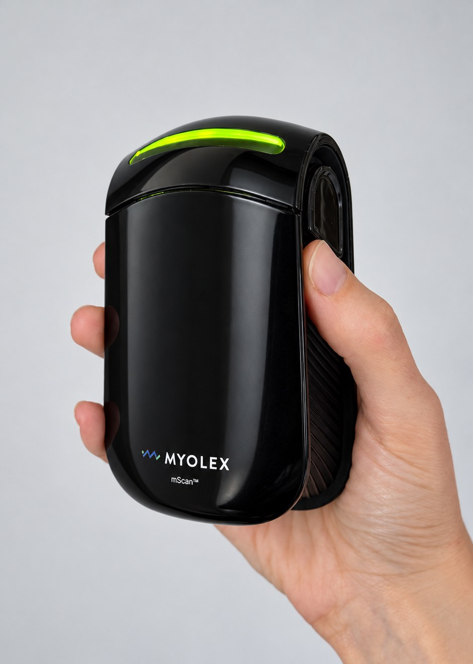
Meet mScan
A portable handheld instrument for fast, localized muscle measurement, in clinic or at home.
- Handheld measurement
- Portable and designed for localized muscle assessment.
- Multi-frequency scan
- Captures the muscle’s electrical response across a broad frequency range.
- Guided research software
- Supports muscle selection, scanning, and quantitative data review.
Why individual muscles matter
Disease and treatment can affect one muscle differently from another. Current tools leave gaps when a study needs to follow a specific muscle over time.
Whole-body tools
DXA and BIA measure the whole body and do not directly characterize individual muscles.
Performance tests
Strength and function tests depend on effort and cooperation, and are not possible in every population.
Imaging
MRI and CT are detailed but costly, not always accessible, and difficult to repeat frequently.

The mScan workflow
A portable handheld instrument with guided software, for use in clinic or at home.
- 1
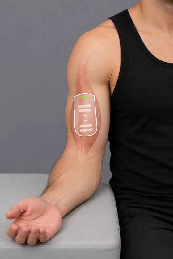
Select a muscle
The app guides placement on the chosen muscle site.
- 2
Collect a scan
Hold mScan on the skin while it runs a multi-frequency sweep in seconds.
- 3
Review measurements
Resistance, reactance, and phase are displayed across frequency for each scan.
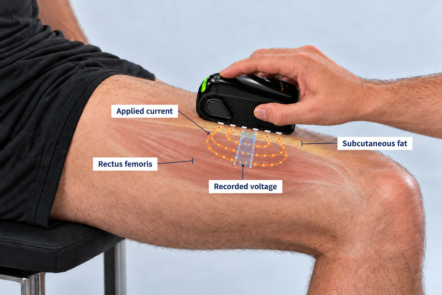
How EIM works
mScan rests on the skin over a muscle and sends a weak, painless current through it. How the muscle resists and stores that current reflects its composition and structure.
- 1
Send current. A low, imperceptible current passes through the skin into the muscle.
- 2
Measure. Electrodes record the voltage at 41 frequencies.
- 3
Calculate impedance. Resistance, reactance and phase per frequency. Changes in muscle structure and composition can alter EIM measurements
Three core readouts from one scan
At each frequency, mScan records the muscle’s impedance, summarized as resistance, reactance, and phase.
Resistance, reactance, and phase defined
- Resistance Ω
- How readily current passes through intra- and extracellular ionic pathways. Varies with tissue geometry, fluid content, and composition.
- Reactance Ω
- Capacitive behavior from cell membranes and tissue interfaces. Sensitive to changes in muscle structure.
- Phase angle °
- Derived from resistance and reactance; summarizes their relationship at each frequency.
Research applications
Neuromuscular disease
Studied in ALS, Duchenne muscular dystrophy, and other neuromuscular conditions.
Longitudinal monitoring
Repeated, effort-independent measurements of the same muscle across a study.
Aging
Participants have collected measurements at home in proof-of-concept research.
GLP-1 and weight loss Exploratory
Early work on whether site-specific EIM can help characterize muscle changes during GLP-1–associated weight loss.
Evidence
EIM was pioneered by Myolex founder Seward Rutkove, MD , at Beth Israel Deaconess Medical Center and Harvard Medical School.

Seward B. Rutkove, MD
Founder, Myolex
Founder of Myolex, Dr. Rutkove is the Nancy Lurie Marks Chair of the Department of Neurology at Beth Israel Deaconess Medical Center and Professor of Neurology at Harvard Medical School. He received his medical degree from Columbia University’s College of Physicians and Surgeons, and his bachelor’s degree, magna cum laude, from Cornell University, College of Arts and Sciences.
Dr. Rutkove’s research focuses on the application of innovative techniques for the assessment of neuromuscular disease with an emphasis on electrical impedance and ultrasound methodologies. His work has been supported by numerous grants from the National Institutes of Health, NASA, and multiple foundations and has resulted in well over 170 peer-reviewed publications.
In 2011, he was awarded the Biomarker Challenge Prize from the non-profit foundation Prize4Life, Inc for his work demonstrating that electrical impedance measurements of muscle could speed clinical therapeutic studies in amyotrophic lateral sclerosis.
FSHD
Detects progression over 12 to 24 months
In 157 adults with facioscapulohumeral muscular dystrophy across 8 sites, EIM phase showed excellent test–retest reliability, correlated strongly with clinical function, and declined measurably by 12 months. Annals of Neurology, 2026. Read paper ↗
Screening
Distinguishes neuromuscular disease
Using mScan on nine muscles in 119 adults and 111 children, machine learning-enhanced EIM identified neuromuscular disease with 84% accuracy in adults and 93% in children. Annals of Biomedical Engineering, 2026. Read paper ↗
Aging
Predicts grip strength in older adults
In adults with and without sarcopenia, multi-frequency forearm EIM combined with machine learning explained 48% of the variance in hand grip strength. Muscle & Nerve, 2025. Read paper ↗
Frequently asked questions
What does mScan measure?
mScan measures the electrical impedance of an individual muscle through the skin: resistance, reactance, and phase, across a range of frequencies. These values reflect the muscle’s composition and structure.
Which muscles can be assessed?
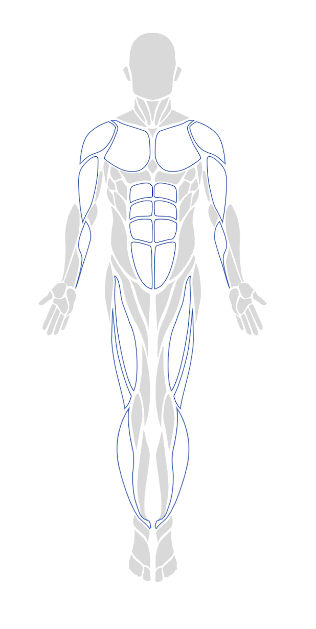
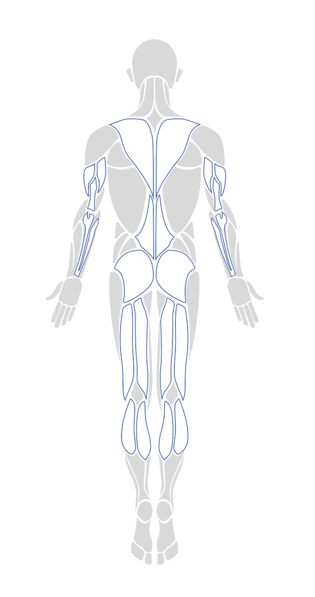
The guided app includes placement sites for muscles of the arms, trunk, and legs, including the deltoid, biceps, triceps, wrist flexors and extensors, rectus abdominis, trapezius, lumbar paraspinals, gluteus, quadriceps, hamstrings, tibialis anterior, gastrocnemius, and soleus.
What does a scanning session involve?
The operator selects a muscle in the app, places mScan on the skin at the guided site, and holds it in place while the multi-frequency scan runs in seconds. No needles or muscle contraction are required, and sessions can be run in clinic or at home.
How can we discuss using it in a study?
Use the form below or email info@myolex.com. Tell us about your population, muscles of interest, and timeline, and our team will follow up.
Bring individual muscle measurement into your study.
mScan is available for investigational use in clinical research.
- info@myolex.com
- Phone
- 888.382.8824
- Office
- 1309 Beacon St, Suite 300
Brookline, MA 02446
Regulatory status
The Myolex mScan system is pending FDA clearance. It is available for use in IRB-approved clinical research. It is not currently cleared for diagnostic use, and nothing on this page should be read as a diagnostic claim.
Thank you
We’ve received your message and will be in touch.